U.S. researcher among 3 awarded Nobel Prize in chemistry for developments in electron microscopyÂ

The Nobel Prize in Chemistry was awarded to three researchers for their work on electron microscopy. (Oct. 4, 2017)
Reporting from STOCKHOLM â Three scientists who developed new ways to capture the clearest snapshots of lifeâs complex molecules have been awarded the Nobel Prize in chemistry.
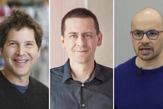
Jacques Dubochet of the University of Lausanne in Switzerland, Joachim Frank of Columbia University in New York and Richard Henderson of the MRC Laboratory of Molecular Biology in Cambridge share the $1.1-million prize for their contributions to cryo-electron microscopy. This technique has allowed researchers to study all kinds of biomolecules in unprecedented detail, from the proteins in our bodies to the viruses that attack us. And scientists say it will help them make breakthroughs in fighting diseases, developing pharmaceuticals and even improving agriculture.
âNow we can see the intricate details of the biomolecules in every corner of our cells, every drop of our body fluids,â Sara Snogerup Linse, chair of the Nobel Committee for Chemistry, said during Wednesdayâs briefing at the Royal Swedish Academy of Sciences in Stockholm. âWe can understand how they are built, and how they act, and how they work together in large communities. We are facing a revolution in biochemistry.â
Frank was roused in the early morning before the announcement but told the academyâs secretary general, GĂśran K. Hansson, that he âdidnât mind.â
âI was fully overwhelmed,â Frank said of the news, adding that heâd thought the chances of winning a Nobel were âminuscule.â
âThere are so many other innovations ⌠that happen almost every day,â he said.
Thereâs a reason images are so important in chemistry, said Allison Campbell, president of the American Chemical Society: They provide information a mere chemical formula cannot.
âA picture gets you right to the heart of the matter,â she said. âI can tell you that my house was 2,000 square feet and had five rooms, but you wouldnât really know how big those rooms were, or what their orientation is relative to each other, whether my house was a two-story or a ranch. But if I give you a picture or a blueprint, you would know exactly how things were put together, organized, related to one another. Thatâs the same thing here.â
Electron microscopes allow scientists to see structures much smaller than is possible with traditional microscopes because the wavelength of an electron is so much smaller than that of light. Youâd think that would make it an ideal tool for studying the structures of life â cells, and the molecules that make them.
But for decades, this was considered impossible. Thatâs because electron microscopes require samples to be placed in a vacuum â which would suck all the water out of tissue or biological structures, thus destroying or deforming them. And the electron beam is strong enough to burn delicate biomolecules, causing irreparable damage.
(Another technique, X-ray crystallography, works for orderly, crystalline biomolecules like DNA â but it isnât helpful for not-so-organized ones.)
Dubochet, Frank and Henderson, each in their own ways, managed to chip away at this problem. Henderson showed that three-dimensional images were possible with electron microscopy; Frank created the software that could sew together three-dimensional images of complex biomolecules; and Dubochet devised an ingenious method to keep samples picture-perfect.
In 1990, Henderson used an electron microscope to create a 3-D image of bacteriorhodopsin, a purple-hued protein that captures solar energy for photosynthetic bacteria. Hendersonâs work succeeded in part because of bacteriorhodopsinâs special features: The proteins could be protected from vacuum by a glucose solution, which isnât an option for all biomolecules. And bacteriorhodopsin molecules packed themselves in a regular way that made it easier to discern their shape â which not all biomolecules do.
Still, Henderson showed that it really was possible to make 3-D images of biomolecules using electron microscopy â and he continued to chip away at the problem over the course of his career.
Meanwhile, from 1975 to 1986, Frank had been working on a way to process images that would allow scientists to pull more information out of them.
When you use an electron microscope on a bunch of proteins, lying any old way, it leaves a shadow-like image. Depending on each proteinâs orientation, that shadow will look a little different. (Think of what your shadow looks like when the sun is shining on your side and when itâs shining from behind you: The first shows your profile, while the second shows your outline from the front. Theyâre of the same person, but from different angles.)
Frank grouped similar images (clearly taken from the same angle) together to create a much higher resolution than the individual snapshots. He then took all those âaveragedâ images and combined them to build a three-dimensional structure.
But to get a really clear picture of what the atomic structure of these biomolecules looked like, scientists believed theyâd have to freeze them. Hereâs the problem: Lifeâs molecules operate in water, so it would make sense to image these molecules in a watery solution. But when water is frozen to ice, it forms a crystalline solid, which blocks the sample from view.
Dubochet found a way around this problem by freezing the watery sample so fast that it did not have time to form a crystal. The molecules remained disordered as they do in glass â hence the term âvitrificationâ (from Latin vitrum, or glass).
Scientists lined up to study Dubochetâs new process, which involved a catapult-like device hurling a sample into ethane that had been cooled by nitrogen to -196 degrees Celsius.
âEveryone working in this field traveled to his lab in order to learn the technique,â Peter Brzezinski, member of the Nobel Committee for Chemistry, said at the briefing in Stockholm.
Before Dubochetâs technique, these complex proteins looked more or less like lumpy, opaque blobs. Thanks in part to his vitrification method, researchers now can see the atomic structure within them.
Cryo-electron microscopy already is being used to fight diseases and improve drugs, scientists said. For example, researchers used this technique to create a 3D image of the Zika virus, which is helping them devise strategies to fight it. Theyâre also learning more about circadian rhythm, which regulates our sleep-wake cycle, by studying the protein that governs it.
The next step, scientists said, will be to start âfilmingâ biomolecule behavior.
âThink about taking a movie rather than a bunch of static images on the time scale that molecules move and perform,â Campbell said. âI think thatâs the next big leap in this area.â
Follow @aminawrite on Twitter for more science news and âlikeâ Los Angeles Times Science & Health on Facebook.
MORE IN SCIENCE
2 Caltech scientists share Nobel Prize in physics for gravitational wave discoveries
Flush with a new Nobel, the LIGO team parties at Caltech
3 Americans win Nobel Prize in medicine for uncovering the science behind our biological clocks
UPDATES:
2:55 p.m.: This article was updated throughout with additional detail.
3:55 a.m.: This article was updated with additional background on the prize.
This article was originally published at 2:50 a.m.


